what is electrophoresis?
Electrophoresis is the movement of particles/molecules under a spatially uniform electric field in a buffer.
When charged molecules are placed in an electric field, they migrate toward the +ve or -ve pole according to the charge in the molecule. The rate of migration of the molecules depends on the strength of the electric field, net charge, size, and shape of the molecule, and also on the ionic bond strength, viscosity, and temperature of the medium(in which the molecules are moving). Electrophoresis is the most widely used technique in molecular biology and biochemistry.
In this technique, a gel is prepared of agarose for DNA and polyacrylamide for protein molecules. The gel is immersed with an electrophoresis buffer to provide ions to carry a current and some buffer to maintain pH at a relatively constant value.
Electrophoresis can be divided into two parts based on the working mechanism.
-
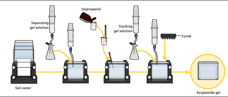
Vertical gel electrophoresis:
- This is used for the separation of protein molecules and is also known as polyacrylamide gel electrophoresis.
-
Horizontal Gel electrophoresis:
- This is used for the separation of nucleic acids and is also known as agarose gel electrophoresis.
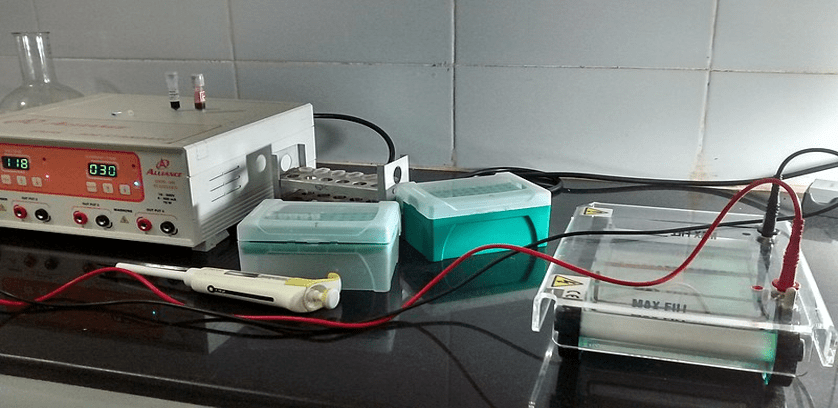
Figure :Agarose gel electrophoresis DNA(ImageRef 3)
Basic Requirements
Support, Gel casting glass slab/trays, electrophoresis unit, power source, sample
DNA GEL ELECTROPHORESIS
DNA gel electrophoresis is the technique for separating DNA based on size and charge(-ve) to visualize and purify. In this technique, we use an electric field to move the -ve charged DNA towards a positively charged electrode through an agarose gel matrix. In this matrix, the shorter fragments of DNA move faster than the larger fragments. For the accurate determination of DNA fragments length, we run it on agarose with a DNA ladder (DNA fragments of known lengths ).
If the size of two molecules/fragments is similar they’ll migrate together in the gel.
AGAROSE GEL
Agarose is a linear polysaccharide that is made of the repeating unit of Agarobiose, which is comprised of an alternating unit of galactose and 3,6-anhydrogalactose. Agarose is obtained from seaweed. The concentration of agarose for electrophoresis is between 1-3%.
Procedure
The preparation of the gel is made by dissolving agarose powder in boiling buffer solution(for proper mixing of agarose). The pore size of the gel is inversely proportional to the concentration of agarose. Then the solution is cooled to approx. 55°C was then poured into the casting tray. A com is attached to form the wells in the gel and later on the comb will be removed after the gel gets solidify.
After solidifying the gel, it is placed in a buffer-filled electrophoresis unit which contains a +ve and -ve electrode. The volume of the buffer in maintained 1/3 of the electrophoresis chamber.
Preparation of the Sample and Loading
For the preparation of the sample, loading dyes are mixed with the sample. Loading dye is made at 6X conc. (0.25% bromophenol blue, 30% glycerol and 0.25% xylene cyanol). The most commonly used dyes in this technique for the visualization of the DNA bands contain Ethidium bromide or methylene blue.
Note: Ethidium bromide is a mutagen that must be handled carefully and disposed of according to the guidelines.
After the preparation of the samples, the samples are loaded into the wells of the prepared gel. And the electrodes are connected to the power source. Usually, for the experiment, the 70-80V DC charge is provided. Then leave it to run the DNA fragments towards +ve charge.
Note: Always place the gel in the electrophoresis chamber so that the wells are placed at the -ve electrode.
Visualization of the Fragments
After the completion of the experiment turn off the power source and remove the lid of the chamber. Take the gel from the chamber and drain off the excess buffer. Take the gel to the gel doc to visualize it under UV light.
Factor affecting the mobility of DNA fragments:
- The concentration of agarose: Higher concentrations of agarose facilitate the separation of small DNAs, while low agarose concentrations allow the resolution of larger DNAs.
- Voltage: In the higher voltage the fragments may move faster than normal and in the lower voltage movements of the fragment are slowed.
- Electrophoresis buffer: Most commonly TAE(40mM tris acetate, 1mM EDTA) and TBE(45mM tris borate, 1mM EDTA) are using as running buffer for electrophoresis.
DNA fragments move differently in both the buffers due to differences in ionic strength.
- Effects of ethidium bromide: EtBr is a fluorescent dye that intercalated between the bases of nucleic acids and allows very appropriate exposure of DNA fragments. The binding of EtBr to DNA may alter its mass and rigidity and it might affect the movement of fragments.
References:
- Principles and Practice of Agarose Gel Electrophoresis: EDVOTEK
- Agarose Gel Electrophoresis for the Separation of DNA Fragments.Pei Yun Lee, John Costumbrado, Chih-Yuan Hsu, Yong Hoon Kim J Vis Exp. 2012; (62): 3923.
- https://commons.wikimedia.org/wiki/File:Agarose_gel_electrophoresis_DNA.jpg