- Gram staining is a method used to differentiate two groups of bacteria.
- Based on the cell wall.
- Developed by Hans Christian Gram in the 19th
- This method is used to identify gram +ve gram –ve
- After performing staining the Gram-positive bacterium stain violet and Gram-negative bacterium stain pink.
- This technique of staining can’t define bacteria, used to general identification such as morphology.
How does Gram staining work?
- Gram staining is a three-step process:
Staining with crystal violet dye, decolorization, counterstaining
- Due to the difference of (thick and thin layer) cell wall, the cells are stained violet and red.
- The cell wall of the bacterium is made up of peptidoglycan.
Steps of Gram staining:
- Stain the cell using crystal violet dye.
- Add Gram’s iodine. (to form a complex between dye and iodine CV-I complex)
- Add decolorizer (ethyl alcohol/acetone).
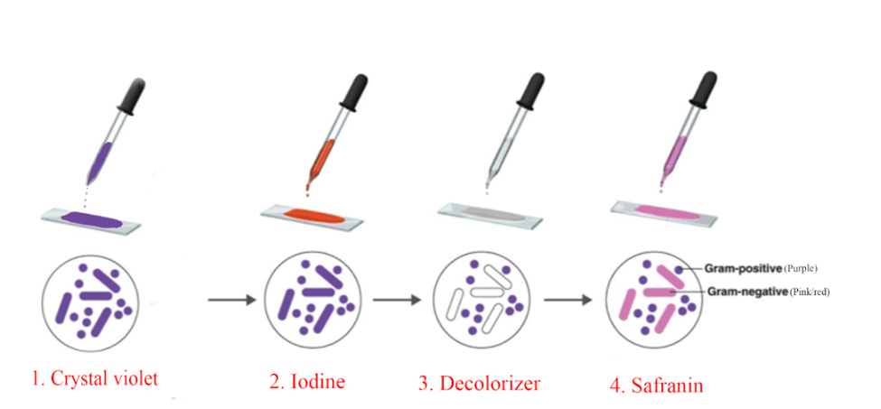
- Adding decolorizer dehydrates the cell.
- After adding decolorizer, the CV-I complex trapped between gram-positive cell walls because it is very thick, and the CV-I complex in the case of gram-negative is dissolved with water.
- Add counterstain (safranin)
- Adding safranin doesn’t disturb the gram-positive cell it only affects gram-negative cells and stain red.
- Contain a single membrane (monoderm) which is surrounded by a thick layer of peptidoglycan.
- Stain purple after gram staining.
- Teichoic acids are present on the peptidoglycan.
- A thin layer of peptidoglycan is present.
- LPS present (lipopolysaccharide)1
Difference between Gram Positive and Gram Negative bacteria:
| Property | Gram-positive | Gram-negative |
|
Around 40-80nmnm | Thinner than gram-positive,30nm |
|
Stain blue(violet/purple) color in gram staining | Stain pink in gram staining |
|
Absent | present |
|
Cell wall contains teichoic acid (-ve charged) | Teichoic acids are absent |
|
Small and single | Two periplasmic space are present |
|
Low | High (because of LPS) |
|
Thick and multilayered | Thin and single-layered |
|
Absent | present |
|
2 rings | 4 rings |
|
Exotoxin | Endo or exotoxin |
|
Some belong to the pathogenic group | Most of them are pathogens |
|
Absent | Present |
|
More prominent | Less prominent |
|
Cocci or rods (spore-forming) | Rods (non-spore forming) |
|
More susceptible | More resistant to drugs |
|
Staphylococcus, streptococcus… | Escherichia, salmonella… |
|
High | Sensitive/low |
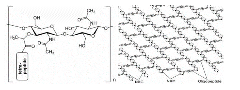
Peptidoglycan:
- Peptidoglycan is also known as
- PG is a polymer of sugars and amino acids.
- PG forms a mesh-like structure outside the plasma membrane.
- The sugar molecules (of Murein) of alternating residues NAM and NAG linked by β-1,4 (glycosidic bond)
- NAM (N-acetylglucosamine), NAG (N-acetylmuramic acid).
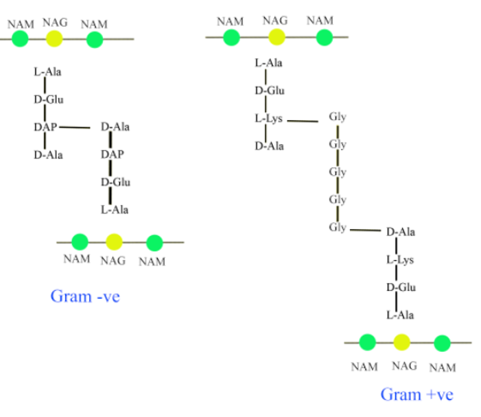
- NAM is a peptide chain of 3-5 AA (amino acids).
- In the peptidoglycan of gram-positive L-Lys is present and in gram-negative in place of L-Lys DAP (diaminopimelic acid) is present.
Question:
Can we heat fix the cell in this method of staining? Explain why?